How to brief and check a cell signaling pathway diagram
A cell signaling diagram communicates relationships. An arrow in the wrong direction can change the meaning even when the illustration looks polished. Start with an explicit relationship list, then compare the draft with that list.
This example is fictional. L, R, K, T, G and I are teaching labels, not claims about a real pathway. Abfig generated the image from our brief; it did not query a biological database or validate a mechanism.
1. Separate components from relationships
Our components are an extracellular ligand L, membrane receptor R, cytoplasmic kinase K, transcription factor T, gene G in the nucleus and cytoplasmic inhibitor I.
| Source | Relationship | Target |
|---|---|---|
| L | Activates | R |
| R | Activates | K |
| K | Activates | T |
| T | Enters the nucleus and activates | G |
| I | Inhibits | K |
This is more precise than asking for a signaling pathway. Use names and relationships supported by your own sources. Mark uncertain or indirect relationships explicitly instead of letting a visual model invent their meaning.
2. Give the diagram a clear scope
The pathway diagram maker accepts a written brief. Our example asks for short labels, named compartments, activation arrows and an inhibition line with a blunt endpoint at K. It requests a legend and prohibits additional components or experimental results.
Start with one cascade or branch. A smaller diagram is easier to compare with the relationship list. If you already have a layout, you can start from a scientific sketch. Do not silently simplify a real network just to make an attractive picture; explain what you have omitted in your accompanying text.
3. Compare the result with the brief
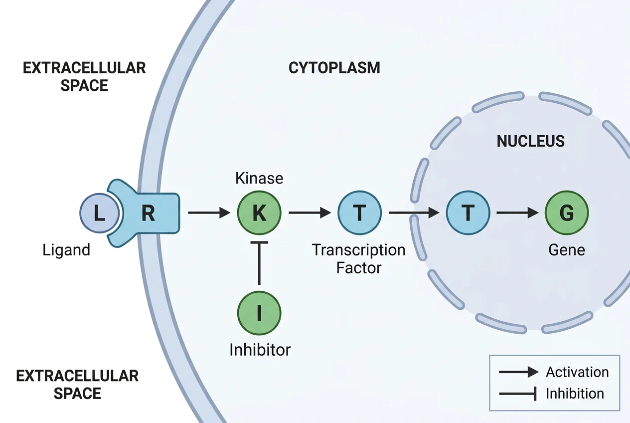
The six requested components are present. The draft shows the main R–K–T–G direction, T at the nucleus boundary and an inhibition bar ending near K.
There are also deviations: the requested cell membrane label is missing, and extracellular space is labeled twice. Correct these before using the figure. Ligand binding is represented by proximity to the receptor; decide whether that representation is clear enough for your audience.
This single result demonstrates a drafting workflow. It does not establish reliable arrow accuracy across pathways or scientific correctness. In a real mechanism, check every edge against the cited source, including its direction, sign and compartment.
4. Revise and review again
Refine uses the current image as a reference and redraws it from an instruction. Selected-area editing redraws a region. Both may change details you intended to preserve. After a revision, check the full relationship list again, not only the edited area.
The scientific figure generator explains the broader conceptual scope. Abfig does not provide native SVG objects, reaction balancing or pathway-database exports.
5. Check readability and delivery
Export RGB PNG or TIFF under your access entitlement and inspect the saved file at its intended size. The DPI checker helps compare dimensions and resolution. Higher DPI cannot correct a missing label or reversed arrow. Before publishing, separately check the venue’s current AI-image policy and disclosure requirements.
Turn this guidance into a visual draft
Use Abfig to explore a layout and communication draft. Check every scientific detail and the target journal's current policy before using it.
Table of Contents
Author
Categories
Tags
More Posts

Does PLOS ONE Require a Graphical Abstract? Current Guidance
PLOS ONE does not describe a separate graphical abstract submission. Learn what its optional Striking Image is and where to verify current figure rules.
Nature Graphical Abstracts: Journal-Specific Requirements
Nature Portfolio has no single graphical-abstract rule. Compare Nature Communications and Nature Chemical Biology, then check the exact title.
Graphical Abstract Layout Structures: Which One Fits Your Research
Compare three useful layout structures—flow, central figure, and comparison—and choose one that matches the scientific story.